Male sex cells - spermatozoa - are movable cells with a length of about 70 microns. The sperm is distinguished by a thickened, rounded head and a thin long tail. The head contains the kernel, in front of which is the structure that called the Acrosoma. Acrosoma has a set of enzymes that are capable of dissolving the egg shell when fertilization. The spermatozoid tail contains contractile elements (fibril beams), ensuring the movement of the spermatozoa. When passing through the seed-handing paths, men to spermatozoa are added liquid secrets of sex glands - seed bubbles, prostate and bulburetral glands. As a result, a liquid medium is formed - the sperm in which
sperm sperm. Life expectancy and fertilizing ability of human spermatozoa ranges from several hours to two days.
Women's sex cells - egg cells - have a rounded shape and large dimensions (up to 150 μm in diameter). Each egg cell contains a kernel and a large amount of cytoplasm, in which in addition to cellular organelles there are protein-lipid inclusions (yolks), glycogen necessary for powering the egg. In a person, due to the fact that the germ develops intrauterine and eats at the expense of the body of the mother, the need to create large reserves of yolk in the egg. Therefore, in the cytoplasm of eggs, women contain a very small amount of yellow and carbohydrate inclusions. Its supply of nutrients eggs usually consumes within 12-24 hours after ovulation. If the fertilization has not come, the egg cell is dying after this time.
Human egg has two covering her shells. Knutrice is a cytlemma, which is a cytoplasmic membrane of an egg cell. Outside from the cytlemma is located a layer of so-called follicular cells that protect the egg and having a hormone-forming function.
Follicular cells form and distinguish female sex hormones - estrogens.
The development of genital cells occurs in germ glanes. The spermatozoa is formed in the testicles of men, egg cells - in the ovaries of a woman. The cycle of the development of spermatozoa is called spermatogenesis. Egg development cycle - Ovogenesis (from Lat. Sperma - Seed, Ovum - Egg, Genesis - Development).
The development of genital cells is finisted by their readiness to fertilization (merger) and the further formation of the embryo. The preparedness of the male and female genital cells to fertilization is not only in the acquisition of specific features of the structure. In the process of spermatogenesis and evogenesis, complex transformations of precursors and young sex cells occur. As a result of a peculiar cell division - MEIOS - in sperm and egg cells there is a decrease (reduction) of the amount of chromosome from the double (diploid) to the haploid (single) set. Instead of usual for all other human cells, the diploid set in the form 46 chromosomes in each cell (spermatozoa, egg) is available according to a single set - 23 chromosome.
Spermatogenesis. The spermatozoa is formed in humans during the entire active period of life of a man. The duration of the development and formation of mature spermatozoa from their previous
sverders - spermatogoniov is about 70-75 days. This process takes place in the walls of the convolves seed tube eggs. Initially, spermatogonia, the total number of which in one egg reaches 1 billion, intensively multiply, are divided by mitotic path. This increases the number of new cells - spermatogonium. In the future, part of the spermatogonium retains the ability to divide and supports the population. Other spermatogonia is added twice in the form of Maiz. As a result, from each such spermatogonium, which has a diploid - double set (.p \u003d 46) chromosomes, 4 sperms are formed. Each of these stems received a haploid (single) set of chromosomes (n \u003d 23). Sperm gradually turn into spermatozoa. This complex process is characterized by the restructuring of spermage structures, which are lengthened and in which a thickened head and a thin long tail is formed. The top of the sperm head is formed by a compacted cart - acrosoma containing enzymes that, when meeting with a female genital cell (egg) destroy its shell. This is important for the penetration of spermatozoa inside the egg. With underdevelopment or absence of acrosoma, the spermatozoa is not able to penetrate the egg and fertilize it.
The formed spermatozoa goes into the clearance of the seed tube tubers. Together with the liquid released by the walls of the tubules, the spermatozoa is gradually moving towards the appendage of the egg, which also serves as a tank for spermatozoa. The amount of spermatozoa generated is huge. 1 ml of sperm contains up to 100 million spermatozoa. These are movable cells, the speed of their promotion through the tube canals is about 3.5 mm per minute. In the female genitals, sperm retains vitality for 1-2 days. They move towards the egg, which is caused by Chemotaxis.
Ovogenesis. Egg, unlike male genital cells, multiply, increase in the amount of embryos, female fruits, i.e. when the fruit is still in the womb. At the same time, the so-called primordial follicles are formed, located in the deep layers of the ovarian cortical substance. Each such a primordial follicle contains a female sex cell - a rogue, surrounded by one layer of follicular cells. Ovogonia is repeatedly mothotically divided, turning into first-order oocytes (primary octivi), which are preserved in the ovary of the girls up to its puberty. By the beginning of puberty in the ovaries there are about 300 thousand primary oocytes. Primary oocytes, with a diameter of about 30 microns each, together with its surrounding two
follicular epithelium cell layers form primary follicles.
In girls during puberty and in half-plant women, most primary oocytes dies. During the life of a woman, only 400-500 eggs ripen.
In the process of ripening, the primary ochocyte passes the stage of mezzani. As a result of meiotic division, a secondary ovocyte is formed, having a single (haploid) set chromosome (/ 7 \u003d 23), and a small, so-called polar caller with the same (and \u003d 23) chromosome. In this case, the primary follicles turn into secondary follicles. Inside such follicles, liquid accumulates, and around each secondary ocylate, two shells are formed - cytlemma and a layer of follicular cells. Thus, the secondary follicle turns into a bubble (tertiary) follicle filled with follicular liquid.
The diameter of the mature bubble follicle reaches 1 cm. The half-length woman simultaneously matures 1 or, less often, 2 follicles. The remaining follicles growing at this time are subjected to reverse development - atresia. At the site of the death of such non-seated and dead follicles, structures remained the name of atrethic bodies.
Spermatogenesis, like Ovogenesis, is a phased creation of Games from representatives of different floors. The first process means the ripening of spermatozoa, the second - egg cell.
The quality of genital cells affects the ability of reproduction. If for any reason spermatogenesis is broken, pregnancy will not happen. Below you can find out how both processes are developing, which are similarities, differences, which factors affect their violation.
General information about evogenesis and spermatogenesis
The most important planet from the functions of all creatures is reproduction, the continuation of a kind. During the self-reproduction, the born organism inherits the genetic material from the parents of the robust, from which half will give to descendants, the following generations and it is thanks to this function that human race continues, and everything begins with merging the genital cells produced by a female, male organism.

To make a merger, we need 2 processes that biologists are called spermatogenesis, evogenesis. Considering both processes, it is necessary to note the unifying similarities. Stages are presented below:
- reproduction. At this stage, Hameta is beginning to share mitosis. It is important to remember that spermatozoa is produced in a man all their lives from a puberty age segment, in women - in the embryonic stage;
- height. Gamets during this period of development increase, turning into sperm, oocytes of 1 order. Oocytes are larger, since they accumulate a lot of useful substances necessary to embryo;
- maturation. At this stage, spermatocytes are detected, 2 order oocytes are revealed, then they are ripening to mature eggs.
Differences and similarity of processes
The comparative characteristics of each of the processes indicate a mass of similarities and the presence of differences, features of each.
- Gametogenesis passes in gradually, including breeding, increase in size, ripening of genital heights. For spermatogenesis, the formation step is considered a distinctive stage - an additional stage, during which sperm is obtained by the form peculiar to them, the movement device.
- The difference in the number of cells - from 1 order spermatocyte is obtained according to the results of 4 cells, from the oocyte of the same order - only one.
- The formation of egg cells occurs cyclically every 21-35 days. As soon as it is dying (menstruation comes), changes in hormonal balance create conditions (push) to generate, ripening other. Moreover, the release of sperm is extended throughout life.
- The number of genital cells will be varied - per day, the male reproductive system in men gives up to 30 million sperms, in women - up to 500 eggs for life.
- Spermatogenesis is subject to external factors, due to the anatomical location of the semennikov.
Features of processes
If we talk about the main characteristics of spermatogenesis, ovogenesis, you need to focus on the number of obtained Games. Maturation of cells in men is aimed at repeated division, which will become a significant amount of sperm. In women, the ripening process gives only 1 Games.
Features are Games tasks. Egging grows, accumulates nutrients that will need an embryo in case of successful fertilization. The main task of sperms is to maintain mobility to overcome the complex path through the female body to the egg, in order to fertilize it.
The dimensions of Games in men, women differ, which is also due to the length of the existence of cells. Women's life cycle longer than spermatozoa.
Against the background of the favorable conditions, the life of a male seed is just a day, but the egg cell remains throughout the development of the fetus, before birth.
Characteristics of Ovogenesis and Spermatogenesis
By apparing the comparative characteristics of both processes, we consider the estimated spermatogenesis, Ovogenesis by:
- localization of education: in semenniki, in ovaries (respectively, sex);
- games: spermatozoa, eggs;
- cell size: 50-54 MK, 120-138 MK;
- activity: Movable, fixed;
- appearance: spoonful with tail, oval;
- the cluster of the nutrient fluid: not accumulate, accumulate;
- the type of division: during mitosis, sperm, oocytes are obtained;
- growth phase: for both processes the nature of the growth of genital cells;
- stage of ripening: Spemidids, ocyats are converted during meiosis;
- the number of phases: 4 and 3, respectively.

Given the comparative characteristics, the differences between the 2 processing processes are of hats by the organisms of both sexes larger than similar signs. This situation is explained by the difference in tasks that are put in front of sperms, women's govetas.
An essential special feature of Ovogenesis, unlike spermatogenesis, is that the number of spermatozoa dying is compensated for every day newly created, but in women the ovarian cycle has limitations.
Already by 40 years, the existing stock of the Ovogoniy is depleted, therefore the beginning of Klimaks comes, infertility. The exact period of each of the women varies, depending on the external, internal conditions.
DNA distribution stages
During the stage, when sex cells are divided, the separation of chromosomes is marked in detail. At the division phase in cell nuclei, DNA molecules are doubled, then chromosomes are redistributed. Such redistribution is considered in stages.

There are 4 stages:
- Latent. It is possible to notice the kernel and twisted thread chromosomes. At the same time, the chromosome of the father and the mother are distant.
- Zigotens. Chromosome parents come into contact, exchange genes.
- Patchyt. Communications in chromosomes are strengthened, twisted with each other.
- Diplotype. At this stage, the circumferential chromosomes are doubled, they are divided into 2 pairs.
Summing up said about sperm and evogenesis, it can be noted that the two most important processes are reduced to the production of genital cells. You can find a lot in common in the processes, but there are differences, which is due to the anatomy, the functions of a female, male organism.
Comparing both processes, we conclude that the goal is common to prepare a seed of different organisms to reproduction, the successful conception of the offspring, to continue their own kind. Given that the tasks of male and female seed will differ, their development is somewhat different.
Gametogenesis affects not only a successful conception, but also the health of future generations. Many unfavorable factors are able to negatively affect the goves of women, men, and this is fraught with infertility or conception of the fetus with congenital anomalies.
It is so important to follow health, plan pregnancy, to undergo prophylactic inspections and comply with the recommendations of doctors.
Ministry of Education and Science of the Russian Federation
FGBOU VPO "Penza State University"
Medical Institute
Department of clinical morphology and forensic medicine with a course of oncology.
Course work on discipline
"Histology, cytology, embryology."
"Spermatogenesis and oogenesis. Similarities and differences"
Performed: Art. c. 12L6 Demoleva Own.
Checked: Assistant Yunashina Yu.V.
Introduction
Spermatogenesis
Comparison of spermatogenesis and oogenesis
Conclusion
Bibliography
Introduction
Reproduction or reproduction, inherent in all living beings, the reproduction feature of themselves like. Unlike all other vital functions of the body, the reproduction is not aimed at maintaining the lives of a separate individual, but on the preservation of its genes in the offspring and the continuation of the kind - thereby the preservation of the gene pool of the population, species, family, etc. During evolution, different groups of organisms were formed - in many cases, independently different paths and reproduction strategies, and the fact that these groups survived and exist, proves the effectiveness of various ways to implement this process.
With a gender method of reproduction, the offspring, as a rule, has two parents. Each of the parents produces sex cells. Sex cells, or gametes, have half or a haploid set of chromosomes and arise as a result of meiosis. Thus, Gameta (from Garete. Gamete is a wife, Gametes - a husband) - a mature reproductive cell containing a haploid set composite and capable of filling with a similar cell of the opposite sex to form a zygota, while the number of chromosomes becomes diploid. In the diploid set, each chromosome has a pair (homologous) chromosome. One of the homologous chromosomes is derived from his father, the other is from the mother. Women's goveta is called an egg cell, men's -Sermatozooid. The process of education and the development of weights in the germ wearing is a common name - Gametogenesis. All other cells that do not take direct participation in the formation of Games received the name of somatic cells. Gametogenesis is a wide term that means the phased "creation" of highly specialized cells capable of providing the beginning of a new organism.
Primary genital cells - Hungali are descendants of embryonic totipotent cells present in blastoderma of the embryo during the formation of the primary strip. Appear earlier than the gender and exist independently of it. Then they fall into the rear extraordinary Entoderm, migrating into the waters of the intestine and in its surrounding mesenchym, and then move to the dozen mesenter to the Gonada tab. Prior to the development of the gonada, they are actively moving in the body with fluid currents. Once near the sex glare, Hungal is approaching an amosboid method attracted by the protein nature factor that is distinguished by a gonad. Penetrating into the gland (ovaries in females, the seeds in males), sex cells are located in males in the brain, and in females in the cortex layer gonads. In the future, sex cells of milking ripens are in gonads. Gonads of the embryo initially contain a relatively small number of sediments of their primary genital cells. But hitting the gonads, sex cells begin to share vigorously, and their number increases sharply. Cells are divided mittochically. Mitosis provides transfer to two daughter cells of absolutely identical sets of chromosomes containing hereditary information.
1.Spermatogenesis
In the men's sexual system, spermatogenesis occurs in sex glasses (gonads) represented by the pair body - the testicles performing two important functions: -Genctive (male genital cells); - Endocrine (synthesis of men's sex hormones).
These functions are interconnected, although provided by various structural components of the organ.
Spermatogenesis includes four periods: -Razznost; -rost; - Spring; -Forming.
The period of reproduction. Sperm cells are represented by spermatogonia. These are small rounded diploid cells located on the basal membrane of seed convolutions. There are two types of spermatogonium: A and B. Type A is represented by light and dark slightly flattened cells with a light core. Dark spermatogonia - no longer, resting cells are considered stem; Light spermatogonia - cells divided by mitosis. Some of them maintain a cambial cell population, others - during consecutive divisions become spermatogonia type V. The lattes have a pear shape, a large rounded core and a centrally located nucleus. Spermatogonia is replenished by dividing (full mitosis) of stem cells of the male gonad. At a certain point, the daughter cell (derivative of the stem) is divided incomplete, leaving a bridge binding subsidiaries, and joins spermatogenesis. The syntial bond, on the one hand, provides synchronicity of the existence of a clone cells, from the other (due to mass) - heterogeneity and polymorphism of its cells and thereby high viability. Differentiating banks. In the process of such mitotic divisions, subsidiaries do not fully grow to the initial and minor, and eventually prepare GONKI to join meyosis. The period of mitotic differentiating spermatogenesis is completed with the creation of a "secondary" spermatogonium, and congestives clone cells to the Maoyotic period of spermatogenesis. The cells that ended with division and entered in the period of growth and ripening are called primary sperm (first-order sperm).
Growth period. During the period of meiosis, complex changes of the nuclei are made, preparing the cell to the transition to the haploid state. First order spermatocytes are significantly increased in the amount and become the most large spermatogen cells, the DNA content in the kernels is doubled (2N4C). They are separated from the basal membrane of the tubules and are shifted towards the enlightenment of the canal. First-order spermatocytes immediately enter the episode of the first division of MEIOS, the duration is about 22 days. In the Profaise of Maiza I, spermatocyte is growing, and therefore such cells are also called auxocytes, that is, growing. Thus, the largest spermatogenesis cells are preparing for the first division of maturation sperm I order.
Ripening period. In male individuals, the first reduction division of meyosis is completed by the formation of two second-order spermatocytes, or secondary spermatocytes. These are smaller cells than primary, which are closer to the clearance of the tubules. The second equation division ends with the appearance of 4 haploid cells - sperm.
The formation period (spermogenesis). In this period, there is a conversion of sperm in mature sex cells - spermatozoa (sperm). During the formation period, only structural changes of cells occur, since the chromosomal set does not change them, remaining the haploid. At the beginning of spermatogenesis, the cells are still associated with each other cytoplasmic bridges and continue to remain as part of a sycitial clone. Structural changes in sperm lies in:
chromatin seal (due to the substitution of histones with nonregone proteins), the reduction of the kernel, the acquisition of the pear-shaped form:
an acrosoma formation is a flat membrane bag containing a number of lipic enzymes necessary for fertilization. Acrosoma-derivatives of the Golgi complex forms the initial acrosomal granules, which, merging, form a bubble, adjacent to the future front surface of the nucleus and gradually flatly flashes in the form of a hat;
formation of burning of a distal centriol forming an axonsee of the tail (after moving both centrioles to the rear pole of the core); The proximal centril is located in the pressing of the nuclear shell;
the formation of special elements of the cytoskeleton occurs as the tilt forming and includes the appearance of 9 longitudinally lying segmented columns around the centrioleum (binder department), which are detailed with 9 dense fibers located along the periphery of the microtubule pairs of the axonsem (intermediate department). In the main department, a fibrous vagina formed by the longitudinal collisters connected by ribs is formed;
changing the shape and location of mitochondria, which are from elongated and diffusely scattered through cytoplasm: sperms, become spiralized and focus around dense fibers in the emerging intermediate department, closely adjacent to each other;
removal of excess cytoplasm containing organelles and lipid inclusions, from sperm forming in the form of so-called residual calves, which are allocated to the lumen of the Channel.
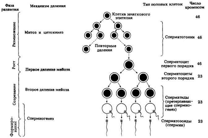
A feature of spermatogenesis is the formation of a functional syncytium combining the clones of spermatogenic cells included in this process. Intercellular communication of spermatogenic cells ensure their synchronous development, nutrient transfer and intercellular exchange of gene expression products. (Fig.1)
Fig.1. Schemes of spermatogenesis processes.
Spermatogenesis in humans lasts 64-74 days, starting during puberty and continuing throughout life. After 50 years, its intensity is significantly reduced. A person is produced daily about 250 million spermatozoa. Spermatogenesis normally flows at a temperature of 3 degrees below the body temperature (temperature in the scrotum). It is inhibited by increasing the temperature (wearing unnecessary warm clothing), cryptorchism (uncomplication of eggs in the scrotum) and pressure on it surrounding the fabrics in the cavity of the peritoneum and inkhan channel.
2.Oogenesis
Eggs - female gamets of animals, higher plants. As a rule, egg cells are haploid cells, but may have a different fairness in polyploid organisms. The human egg has a diameter of about 150 microns.
In the cytoplasm of eggs (ooplasm) contains the inclusion of nutrients - yolk. Eggs are formed as a result of ovogenesis. After fertilization from the fertilized egg (zygotes), the embryo develops. In case of parthenogenesis, the embryo, and then the new organism develops from a non-advocated egg.
The human egg first was first described in 1827 Godr. It strengthened interest in the study of the processes of gaming formation and fertilization.
From spermatozoa, the egg cell differs:
overwhelming real estate;
accordingly, a characteristic more or less spherical shape;
the presence of diverse protective and shells of nutrients sources;
the absence of functional organelles or formations inherent in spermatozoa: tail, specialized mitochondrial complex, acrosomes,;
genetic information (sex chromosomes - XX).
features of education and development, as well as life time;
significantly smaller number in the body (during life in the female organism, about 400 eggs is formed, while spermatozoa in the men's million millions).
feedstock of nutrients for the development of the future embryo, localized in the cytoplasm;
much large size (man's eggs 85,000 times more spermatozoa).
The process of developing female genital cells is called oogenesis. Gonocirats put in the descendant of female sex gonad, and all the further development of female genital cells occurs in it. Having hit the ovary, Hungali becomes oogonia. In this process there is no formation period.
The process of ogenesis consists of three periods: -Razznost; -rost; - Spring.
Unlike spermatogenesis in the ovaries, breeding, growth and partially maturation, ending in the ovitel, occurs. In addition, the end of the second division of meiosis occurs only as a result of fertilization, and therefore the process of ogenesis does not always reach the end.
The period of reproduction. Diploid cells formed from Gonocytes at the 8th week, Oogonia (immature sex cells), mitoto-3-4 months of intrauterine development are repeatedly divided, as a result of which in both ovaries of a person, their number increases, reaching a few hundred thousand. With such a margin of sex cells, a girl is born. New sex cells after birth do not occur and the mass degeneration of genital cells occurs. After the last division in the reproduction period, the cell enters into the epipase of the first division of maturation, and the cell cycle is delayed for a long time. In PROFARE I, MEIOSA occurs to the conjugation of chromosomes, the formation of a synaptonem complex, crosslinker, that is, events that determine all the further processes of meiosis.
Growth period. Oogonia is included in the period of growth. They lose the ability to mitotic division and come into opposition I MEIOS. In PROFARE I, MEIOSA occurs to the conjugation of chromosomes, the formation of a synaptonem complex, crosslinker, that is, events that determine all the further processes of meiosis.
Two phases are isolated: small and large growth. Prior to the occurrence of sexual maturity, the process of small growth is performed when it occurs mainly an increase in the size of the nucleus and the cytoplasmism account of the accumulation of dehytoplasmic substances in the form of a yolk. In the period of great growth, accumulation of nutrient material is accumulated in the cytoplasm, which is brought into the ovary with the blood of the maternal organism. The composition of so-called yolk inclusions includes proteins, fats, leafy-like substances. On the chromosomes of the primary oocyte, a large amount of information and transport RNA is synthesized, as well as a substance of a special composition, located under the plasmolym, forming the cortical layer. The stages are capable, leptoten, zygoten, patchytes, diplotenses occur. At the zigoted stage, the formation of the synaptemal complex and the conjugation of homologous chromosomes begin the formation of meiosis. The synaptonex complex (SC) is a genetically deterministic three-dead protein structure. In Patchithee, the conjugation is completed by the formation of the Bivalent, which reached the reducing reduction of the number of chromosomes. So the primary oocyte occurs, or the first-order oocyte, surrounded at the beginning of the layer of flat follicular cells (the prioritial follicle). The volume of the kernel and cytoplasm increase proportionally and slightly. At the same time, nuclear-cytoplasmic relations are not violated.
Forming primary follicles, in which a brilliant zone appears for the first time, having a view of the structureless oxificial layer between the primary oxygen and the follicular cells of the prismatic form. It performs a number of important functions: -Creatment is a semi-permeable barrier between follicular cells and oocytes; - Enhaps the surface area of \u200b\u200bthe contact between them; - Supplements the species specificity of fertilization; - Complete monosperm fertilization; - Protects an early embryo when moving in sex paths before implantation.
In the first half of great growth, the core and cytoplasm (cytoplasmic growth) increase intensively. "Lamp brushes" and nuclei reaches the most development and actively participate in the synthesis of RNA. In the second half of the largest growth period, visylelogenesis (trophoplastic growth) is carried out. In the kernel there is a decline in RNA synthesis. It is often formed by a carosphere - a special structure with pores consisting of elements of membranes and a synapetemal complex for insulation of diploten chromosomes of the oocyte core from the functional activity of extrachromosomic DNA and nuclei.
At the end of the period of greater growth, "lamp brushes" lose the loops and strongly shortening. The stage of diakinosis occurs, followed by a metaphase plate of the first division of ripening. The nuclei function for a short time or do not develop at all, and the carosphere is early formed. A cite-cytoplasmic relationship is reduced.
At the stage of Diakinosis, the course of meiosis slows down to a complete termination (Maizo block). The meyosis unit in humans is removed with the onset of inheritance. Proofase I can be very long, and a large growth of oocytes capable of ovulation, a person is stretched for decades, that is, on the whole reproductive growth.
With each sex cycle, the oocyte group enters into a period of great growth, but not all of them develop to the end, as most of them stop growing and dying. Only one of them (very rarely several oocytes) proceeds to the next period of oogenesis - maturation.
Ripening period. The accumulation of substances in the cytoplasm of the primary oocyte is completed, and then the remaining phases of the first reduction ripening separation. As a result, two diploid, but unequal sizes of the cell are formed. In one of them, a cage of large sizes, called second-order oocyte, or secondary oocyte, remain almost all the accumulated substances necessary for the further development. Other, small sizes, has very little cytoplasm, and therefore is called reduction, or guilding, tel. The formation of a secondary oocyte in a woman coincides with the moment of ovulation, when after the gap of the mature follicle (grappa of the bubble), which is usually happening on the 14th day of the ovarian - menstrual cycle, the genital cell leaves the follicle. Following this, secondary oocytes of the second order at the metaphase stage of the second division of MEIOS, surrounded by a transparent zone and follicular cells of a radiant crown, falls into the rustic tube funnel. (Fig.2, 3.)
Fig. 2.Cheme processes of oogenesis.
The second division of meiosis is not always completed, but only if the spermatozoic reaches the surface of the oocyte and penetrates it. This division is also uneven, as it leads to the formation of an egg from the secondary oocyte, which preserves all the substances necessary for the development of a new organism, and a new reducing Taurus.
The cyclicality of growth and ripening of genital cells in a half-green female body manifests itself in the fact that 5-20 ocytes are involved in the process of great growth monthly, but only one of them will enter into the ripening phase, and the rest will die in the process of atresia follicles. At the 5-bd decades with the onset of menopause, the development of genital cells is terminated: in the future, it is subjected to degenerative changes and disappear from the ovary.
Fig. 3. Stages of human development:
And - before birth, a small proportion of the priority follicles begins to grow, and these follicles are now called developing. B - after some period of continuous growth, some of the developing follicles accumulate liquid, turning into antral follicles. B - with the onset of sexual maturity. Once a month, the wave of a luteinizing hormone released by the pituitary is encouraged by one antral follicle to ripen: the first order oocyte, which is in this follicle, completes the first division of meiosis, forming a polar caller and turning into second-order oocytes. G is a second order oocyte together with the polar tel and part of the surrounding follicular cells is exempt at the moment when the follicle is broken on the surface of the ovary. The second order oocyte undergoes the second division of MEIOS only if it is fertilized. - Prioritial follicle; II - developing follicle; III - antral follicle; Vi - a large antral follicle (grappes bubble); V - broken follicle; 1 - first-order oocyte, stopped in Proofase I: 2 - follicular cells; 3 - cavity; 4 - Oocyte first order; 5 - lifting the level of LH; 6 - the first-order oocyte completes the first division of MEIOS, turning into a second-order oocyte; 7 - the surface of the ovary; 8 - second-order oocyte; 9 - 1st Polar Taurus.
Ovogenesis occurs with the constant interaction of developing sex cells with epithelial in the composition of follicles.
Comparison of spermatogenesis and oogenesis
Oogenesis has a fundamental similarity with spermatogenesis, oogenesis also passes a number of stages: breeding, growth and ripening.
Despite this, the principal similarity of genetic processes during spermatogenesis and oogenesis, there are significant differences between them.
First, the stage of formation is inherent in spermatogenesis and is absent during oogenesis.
Secondly, the growth stage in oogenesis is longer than when spermatogenesis.
Thirdly, the stage of ripening of oogenesis has its own characteristics, consisting in the unevenness of ripening divisions leading to the release of polar taurus. spermatogenesis oogenesis reproduction
Fourth, in the individuals of the female, the first division of meiosis begins during the period of intrauterine development, first ends at the time of puberty, and on the last one on the eve of Menapuses. Maize boys begins only with the achievement of puberty and persists through the entire sexual maturity of a man.
Fifth, the formation of mature genital cells in women occurs cyclically with a period of about 28 days, while men occur continuously.
Sixth, in contrast to spermatogonium, each excretive as a result of meiosis gives four functionally full spermatozoa, only one egg generation is obtained from the oogonia. After the first division of MEIOS in one daughter cell, most of the cytoplasm departs, and in the second, called the guideline, small. Similarly occurs during the second division of MEIOS. The guing taurus is degenerate.
On-seventh, male and female sex cells are very different in structure and functions: Sperm - small mobile cell, very rich in mitochondria, which supply it energy to move, while the egg cell is the largest cell of the human body (diameter 150 - 200 microns ), containing not only significant reserves of nutrients, but also matrix RNAs that will be used in the early stages of the embryo development. The egg cell is surrounded by feeding it with follicular cells and forms a specialized structure - follicle (grappes of the bubble).
The eighth, the course of the coupperatogenesis is more susceptible to the influence of the factors of the outer cavity rather than the course of Ovogenesis, due to the difference in the location of the genital organs (the semenniks, as a rule, are outside the abdominal cavity).
Fig. 4. Comparison of spermatogenesis and oogenesis.
Conclusion
Sexual reproduction is a significant evolutionary acquisition of organisms. On the other hand, it contributes to the exposure of genes, the emergence of the diversity of organisms and the increase in their competitiveness in continuously changing environmental conditions. Compared to other cells, the game is unique. They provide the transfer of hereditary information between individuals of different generations than retain life in time.
Bibliography
1.Valkov E.I. "General and medical embryology." Tutorial for medical universities. St. Petersburg "Foliant" 2003. Art. 27-34.
With the topic right now, to learn about the possibility of receiving consultation.In this paper, we propose to allocate the difference between the oguenesis from spermatogenesis, talk about these processes themselves. Of course, we will not leave without attention and sex cells, explain in detail their structure and functions.
The reproduction is the main appointment of all the living beings of our planet, it is it that helps to continue the genus, that is, our planet will never empty. On the contrary, now the number of living beings, especially for people, is growing in an egg and spermatozoa - these are women and men, respectively. It is our article that will be devoted to them. Under the Gametogenis is understood as the process of formation of genital cells. If we are talking about sperm, it has the name spermatogenesis, if about egg cells, then evogenesis. All this in more detail you will learn later.
Gametogenesis
Ovogenesis and spermatogenesis, the difference between which is non-critical, similar to many of their own features, can be called one common term - "Gametogenesis". Now about it a little more.
To begin with, we will analyze the very concept, we can allocate two words: "Gameta" and "Genez", the last from the Greek can be translated as "origin". That is, the term "Gametogenesis" denotes the "origin of Games." Gamets are sex cells, in men - spermatozoa, in women - egg cells. Gametogenesis itself can also be divided by sexuality: Gametogenesis, occurring in the body of a man, has the name spermatogenesis, and in the body of a woman - Ovogenesis. But we come to the first difference between these processes. Ovogenesis begins even before the birth of the girl, and spermatogenesis is manifested in boys who have achieved a certain age, as a rule, 12-13 years.
Sexual maturity girl

The process of forming eggs, that is, Ovogenesis occurs in the mother's womb, while puberty, or rather its first stage, falls about nine years. We offer a little more consider the feet of puberty:
- 1st step - enhanced growth, there are cases when the girl is gaining more than 10 centimeters in growth, this is considered the norm. As a rule, now girls overtake boys.
- 2nd stage - changing secondary sexual signs. About 12 years old, the girl begins to leave the image of a nasty duckling. Begins to grow the chest, the waist decreases, the hair in the pubic zone and in the armpits are growing. Approximately at this stage, canned eggs are wanted and further developed their own development. Soon the girl learns what monthly.
- The 3rd stage is the final stage. It comes around for about 18 years. Now the girl is considered to be completely formed, it can reproduce healthy offspring.
Floor maturity boy

As we mentioned earlier, the girls have eggs still in the womb, boys are all a little different. Spermatozoa is starting their development only when 12-14 years have reached. Before this stage of puberty, there are also changes:
- increase in the size of the penis;
- increasing the scrotum;
- grow hair on the pubic, in the armpits, on the legs and face.
Upon reaching 12-14 years, the young man can begin to lead a sex life, but it is worth warming his parents, because he can already cause an unwanted early pregnancy of his companion. We now turn directly to the male and female sex cells, consider their structure, stages of formation, similarities and differences.
Egg
Let's start with the female genital cells, the characteristic of the ovogenesis will be considered a little later. First, we propose to consider the structure and function of the egg.

The egg cell is relatively large and fixed cell, its dimensions reach 170 μm, which is much more male genital cells (up to 70 microns). Each of them contains the necessary nutrients, we will see:
- substances needed for protein biosynthesis;
- regulators;
- yolk.
Cells can be divided by the number of yolk:
- on the alcital;
- polycytal;
- mesocitals;
- oligolecital.
From negligible to very large, respectively. If we consider the female egg, it can be attributed to the alecital and isoletive type. That is, there is little yolk in it, which can be explained by the fact that the human germ quickly moves to the hematotrophic type of nutrition. The isolecital type means that the yolk is distributed evenly and the kernel turns out to be in the center.
The egg cell has the following shells:
- cytoplasm;
- protective shell;
- rady crown.
All shells have a protective function, do not give to penetrate the egg cell more than one spermatozoa designed for fertilization. All others are blocked.
Now lay out the functions of the egg:
- ensuring Embryo Energy;
- ensuring in the initial stage of the embryo with nutrients.
Sperm

Everyone knows that spermatozoa is a men's sexy cell, but how is it arranged? We suggest a little disassemble this question. The appearance of Games you can see the photos of this section. The following parts can be distinguished in its structure:
- head;
- neck;
- middle part;
- tail.
Sperm head is filled with a kernel, it is she who carries hereditary information. During fertilization, the egg cell misses it. The floor of the future child depends on the spermatozoa. If he carries a X-chromosome, then there will be a girl if y, then a boy.
The neck is represented in the form of a small narrowing in front of the middle part of the spermatozoa, it is this part that is responsible for the active movement, otherwise fertilization would be impossible.
Before we consider the features of spermatogenesis and ovogenesis, we propose to highlight the basic function of spermatozoa - this is the report of the genetic material to the egg.
Ovogenesis
Let's start with the formation of a female genital cell, we highlight the periods of ogogenesis and characterize each of them. So, phases are distinguished:
- reproduction;
- growth;
- maturation.

Now we can already name the first difference between the spermatogenesis ovogenesis: in the first case there is a birth of female genital cells (egg cells), and in the second - male (spermatozoa). Let's characterize each stage of ovogenesis.
At the stage of breeding, the initial cells (germs) are divided by mitosis available in the parenchyma. Thus, in the cortex layer of the ovaries there is an accumulation of a ovogonium. They accumulate:
- proteins;
- fats;
- glucose.
Now they are very different from their predecessors, the resulting Ovogonia is much larger primarily, but their genetic composition is identical. It is important to note that this process is happening before the girl's birth, that is, in the womb.
The next stage happens shortly before the girl's appearance. This stage is customary to be called growth. Now the division of mitosis occurs, the first order oocytes are formed. Compared to the Ovogonia, they become smaller, but further rapidly increase in size. Now Omocytes are waiting for a slight difficulty, they are in captivity of the granular shell and stop in the development at the stage of the priority follicle. Total cells are approximately two million, but only some of them will receive further development (approximately 450).
The third stage (ripening) comes shortly before the appearance of the first monthly girls. One of the fallen follicles wakes up and continues its development, which stopped at about 12-13 years ago.
Spermatogenesis
We have already identified one difference between the spermatogenesis of spermatogenesis (various sex cells develop), and it is also possible to select the fact that the Ovogenesis begins before the birth of the girl, but the process of spermatogenesis - when reaching 12-14 years. We have already allocated two differences, then we once again denote to consolidate knowledge.
The stages of spermatogenesis are also slightly different from the stages of ovogenesis (the presence of another stage). The following steps of spermatogenesis are distinguished:
- reproduction;
- height;
- maturation;
- formation.
Here, we see the similarity of these processes, the first three stages are the same, only at the stage of ripening the Ovegenesis ends, and the spermatogenesis continues, the formation stage begins.
Difference
You have practically finished reading this article, try ourselves - name the differences in spermatogenesis and ovogenesis. Now check how you coped with the task.
The first difference between Ovogenesis from spermatogenesis is a different start time of the process. In girls, the process of gametogenesis begins during the intrauterine development, boys in 12-14 years old.
The second difference - in the process of ovogenesis develops egg cells, and spermatogenesis - spermatozoa.
The third difference is the lifespan of sex cells. The sperm lives from a hundred to one hundred and ten days (if you take into account the whole process of spermatogenesis, about 74 days), the female sex cell is the result of a long expectation in the ovary (from 12 to 40 years).
Fourth difference - quantity. In all life, many hundreds of billions of spermatozoa come out for light, and completely all stages of ovogenesis are approximately 450 eggs.
Similarities
We during the writing of the article allocated both similarities and differences in spermatogenesis and ovogenesis. Now again we generalize, we denote in the form of a list of similarity of these processes.
- Three common stages (reproduction, growth, maturation).
- Sperm and oocytes are formed by mitosis.
- The stage of growth and in evogenesis, and in spermatogenesis is characterized by an increase in Games.
- Stage of ripening occurs by meyosis.
Describe spermatogenesis, ovogenesis and structure of genital cells.
Spermatogenesis is the process of forming spermatozoa. Conditionally spermatogenesis is divided into several phases.
- Phase reproduction. The diploid cells of the semenist are repeatedly divided by mitosis.
- Growth phase. Accompanied by an increase in the volume of cytoplasm cells, the accumulation of a number of substances for further divisions. In the formation of male sex cells, the growth is weakly expressed. In the cell growth phase, the name of the schumocytes I of order is obtained.
- Phase ripening. During this period, cells are divided by meiosis. As a result of the first division of MEIOS from one sperm of the first order, the formation of two second-order spermatocytes occurs, each of which, after the second division of MEIOS, forms two sperm. As a result, 4 sperms are formed.
- Phase of formation. During this period, the sperm, characteristic of spermatozoa, acquire sperm. Men's genital cells are much smaller egg cells. They have different animals, they have a different form, but most of them have a head and tail. Thanks to the oscillations of the tail, the spermatozoa is actively moving. The kernel decreases and moves to the head, most of the cytoplasm disappears. Next to the core is the Golgji complex, which is involved in dissolving the egg shell when fertilization. Mitochondria is concentrated at the base of the tail and provide energy to its movement.
Ovogenesis - the formation of female genital cells, is based on the same scheme, but with some differences.
- Phase reproduction. There is a multiple division of the cells of the walls of the ovary and the formation of diploid cells - the ovogonium.
- Growth phase. For female sex cells, this period is long and well expressed. The sizes of cells during this period are very much increased. Idocytes I are formed, they accumulate a large amount of nutrients.
- Phase ripening. In case of evogenesis, has its own characteristics. The protopase of the first meiotic division is carried out in the embryonic period, and the remaining phases of the first and second meiotic divisions occur already after puberty. Every month in one of the ovaries, a female woman ripens one egg. At the same time, the first division of meyosis is completed, a large ovocyte II of order and a small first polar (reduction) Taurus, which come into the second division of MEIOSA are completed.
At the metaphase stage of the second meoyotic division, the second order ovary comes out of the ovary into the abdominal cavity (ovulating), then falls into the ovage. Further ripening it is possible after merging with sperm. If fertilization does not occur, then the ovocyte II of the order dies and is excreted from the body. In case of existence, it completes the second meiotic division, forming a mature egg cell - whether and the second polar caller. Polar Taurus no role in evogenesis play and eventually die. Thus, as a result of the ripening phase from each diploid cell, haploid cells are formed - one egg cell and three polar vents.
- The formation phase for eggs is not expressed.
Eggs of different organisms differ in their structure and sizes. In mammals, they have a diameter of 60-2000 μm, the ostrich has several centimeters. The shape of the egg is usually rounded, in its cytoplasm there are mitochondria, ribosomes and a large amount of spare nutrients in the form of yellow grains and protein.